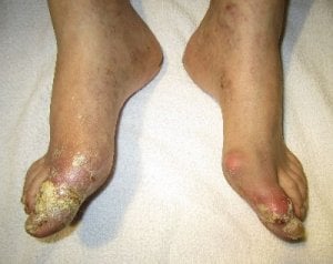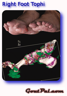
This composite picture is from the public abstract for the first gout study referenced in the Feet Tophi page.
The top photograph shows a large tophus attached directly to the sole of the right foot. You can read more about this type of tumor on the Tophaceous Gout page.
The lower image is a DECT scan which differentiates uric acid deposits, shown here in green. The color is set by the software to provide contrast with natural tissue. Note that, though the patient had not complained of gout pain, uric acid deposits are clearly visible in the toe, foot, and ankle joints. You can find more about this imaging method by searching for DECT in the box above.
Leave Right Foot Tophi to browse more tophi pages
Please give your feedback
Did this page help you? If yes, please consider a small donation. Your donations help keep GoutPal's gout support services free for everyone.
If not, please tell me how I can improve it to help you more.
- YouTube
- The gout forums.