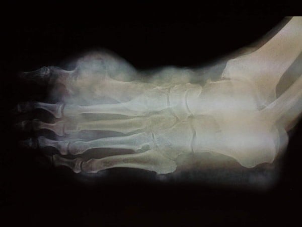
Radiographs of the Gouty Foot.
Total destruction of the first metatarsophalangeal [big toe] joint and soft tissue swelling is shown as is focal involvement of dorsal [upper] and plantar [lower] surface of the foot (panniculitis [inflammation of the fatty layer of tissue under the skin]).
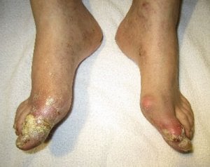
Image from Multiarticular chronic tophaceous gout with severe and multiple ulcerations: a case report
This is the second of three images in Left Foot Tophi Study. Read the report, then do something to make sure you never risk losing your foot like this.
Doctors considered amputating the foot after examining the mess in the previous photograph, and this x-ray showing advanced joint damage from gout. Fortunately, surgeons saved the foot, but do you really want to risk this when you reach 75?
Get uric acid safe today. Just ask in the forums if you are not sure how.
Leave Left Foot Tophi to browse other tophi pages
From: J Med Case Reports. 2011; 5: 397.
Published online 2011 August 19. doi: 10.1186/1752-1947-5-397
Copyright ©2011 Falidas et al; licensee BioMed Central Ltd.
This photograph is distributed under the terms of the Creative Commons Attribution License (creativecommons.org/licenses/by/2.0), which permits unrestricted use, distribution, and reproduction in any medium, provided the original work is properly cited.
Please give your feedback
Did this page help you? If yes, please consider a small donation. Your donations help keep GoutPal's gout support services free for everyone.
If not, please tell me how I can improve it to help you more.
- YouTube
- The gout forums.