Gout in Shoulder X-ray image is my featured image for Gout in Shoulder.
Gout in Shoulder X-ray image details
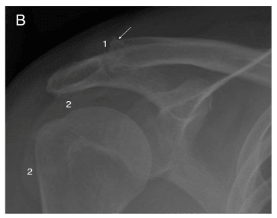
This image is from the Spanish gout case study featured in my Gout in Shoulder article. So, I have translated the original caption using Google Translate.
Simple x-ray of the right shoulder in an anteroposterior projection. In the clavicular portion of the right acromioclavicular joint, a bony erosion in eccentric punches is observed, formed by the replacement of the bone with a less dense tophus (1). Due to its slow and benign growth, the bone reacts by forming a “ring” of sclerosis which, when surrounding the tophus, surpasses even the anatomical limits of the bone forming an elevated border (?). The alteration of the glenohumeral joint is manifested mainly in simple radiology by occupation of the subacromiodeltoid bursa (2).
Leave Gout in Shoulder X-ray image to read Gout in Shoulder.
Please give your feedback
Did this page help you? If yes, please consider a small donation. Your donations help keep GoutPal's gout support services free for everyone.
If not, please tell me how I can improve it to help you more.
- YouTube
- The gout forums.